Pulmonary Consolidation
Definition:
Consolidation of the lung/Pulmonary consolidation
A condition in which lung tissue becomes firm and solid rather than elastic and air-filled because it has accumulated fluids and tissue debris.
Signs of consolidation
- Expansion of the thorax on inspiration is reduced on the affected side (Unilateral reduction in chest expansion)
- Vocal fremitus is increased on the side with consolidation
- Percussion : dullness
- Breath sounds are bronchial
- Possible medium, late, or pan-inspiratory crackles
- Vocal resonance is increased. . Here, the patient's voice (or whisper, as in whispered pectoriloquy) can be heard more clearly when there is consolidation, as opposed to in the healthy lung where speech sounds muffled.
- A pleural rub may be present
Causes of consolidation
When you think of the causes of consolidation, think of 'what is replacing the air in the alveoli'?
Is it pus, edema, blood or tumor cells ?
Is it pus, edema, blood or tumor cells ?

Acute consolidation is seen in:
- Pneumonias (bacterial, mycoplasma, PCP)
- Pulmonary edema due to heart failure or ARDS
- Hemorrhage
- Acute eosinophilic pneumonia
Chronic consolidation is seen in:
- Organizing Pneumonia
- Chronic eosinophilic pneumonia
- Fibrosis in UIP and NSIP
- Bronchoalveolar carcinoma or lymphoma
Most patients who are evaluated with HRCT, will have chronic consolidation, which limits the differential diagnosis.
Radiological appearances common to all lobes are:
1.Abnormal lung opacity2.Increase in the size and number of lung markings3.Loss of clarity of the diaphragm on the AP and/or lateral views4.Loss of clarity of the heart border on the AP and/or lateral views5.Air bronchogram lines6.Loss of the normal darkening inferiorly of the thoracic vertebral bodies on the lateral view7.Opacification of the lung behind the heart shadow or below the diaphragms

1.Abnormal lung opacity------------>
.
.
2.Increase in the size and number of lung markings
.
.
3.Loss of clarity of the diaphragm on the AP and/or lateral views
.
.
4.Loss of clarity of the heart border on the AP and/or lateral views--------->.
5.Air bronchogram lines
.
.
6.Loss of the normal darkening of the thoracic vertebral bodies on the lateral view
.
.
Notes
There is abnormal opacity on the right (arrowed). There is also loss of clarity of the right heart border known as silhouette sign.
Right Lower Lobe (RLL) Consolidation
 •Appears as an area of increased opacity within the RLL
•Some loss of the hemi-diaphragm is commonly seen
 •Loss of right hemi-diaphragm
•Dense opacity in RLL
•Some loss of right heart border
Left Lower Lobe (LLL) Consolidation


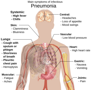
Pneumonia..
An acute or chronic disease marked by inflammation of the lungs and caused by viruses, bacteria, or other microorganisms and sometimes by physical and chemical irritants.
Signs and Symptoms
There is 2 types of Pneumonia:
A. Bronchopneumonia
B. Lobular pneumonia

A. Bronchopneumonia
-is an acute or chronic inflammation of the lungs, in which the alveoli and / or interstitial are affected. Pneumonias are the most common cause of death among infectious diseases. They take the fifth place in the statistics of diseases causing death.
Common causes of pneumonia
~Virus( influenza, ornithosis)
~Gram-Positive Bacteria. These bacteria appear blue on the stain and are the most common organisms that cause pneumonia. They include:
- Streptococcus (S.) pneumoniae (also called pneumococcus), the most common cause of pneumonia. This Gram-positive bacterium causes 20 - 60% of all community-acquired bacterial pneumonia (CAP) in adults. Studies also suggest it causes 13 - 38% of CAP in children.
- Staphylococcus (S.) aureus, the other major Gram-positive bacterium responsible for pneumonia, causes about 2% of CAP and 10 - 15% of hospital-acquired pneumonias. It is the organism most often associated with viral influenza, and can develop about 5 days after the start of flu symptoms. Pneumonia from S. aureus most often occurs in people with weakened immune systems, very young children, hospitalized patients, and drug abusers who use needles. It is uncommon in healthy adults.
- Streptococcus pyogenes or Group A streptococcus.
~Gram-Negative Bacteria. These bacteria stain pink. Gram-negative bacteria commonly cause infections in hospitalized or nursing home patients, children with cystic fibrosis, and people with chronic lung conditions.
- Haemophilus (H.) influenzae is the second most common organism causing community-acquired pneumonia, accounting for 3 - 10% of all cases. It generally occurs in patients with chronic lung disease, older people, and alcoholics.
- Klebsiella (K.) pneumoniae may be responsible for pneumonia in alcoholics and other people who are physically debilitated. It is also associated with recent use of very strong antibiotics.
- Pseudomonas (P.) aeruginosa is a major cause of hospital-acquired pneumonia (nosocomial pneumonia). It is a common cause of pneumonia in patients with chronic or severe lung disease.
- Moraxella (M.) catarrhalis is found in everyone's nose and mouth. Experts have identified this bacterium as an uncommon cause of certain pneumonias, particularly in people with lung problems such as asthma or emphysema.
- Neisseria (N.) meningitidis is one of the most common causes of meningitis (central nervous system infection). The organism has also been reported in pneumonia, particularly in epidemics of military recruits.
- Other Gram-negative bacteria that cause pneumonia include E. coli, proteus (found in damaged lung tissue), enterobacter, and acetinobacter.
~ Aspiration of foreign bodies(food,Vomitus)
~Inhale of suffocating or irritating gases or vapours(benzene,toluene,benzine) or other toxic substance
Symptoms:
- cough
- fever
- dyspnoea
- hyperaemia of the face
- cyanosis of lips
- respiration accelerates to 25-30 per min
- respiratory lagging at affected side
- percussion: lose resonance or completely dull
- ausculatation : vesiculobronchial or bronchial breathing
- Dry and moist rales frequent , but consonating moist rales and crepitation that heard over limited part of chest are especially informative
X-Ray examination
- focal inflammation at least 1-2cm in diameter
Sputum test
-mucopurulent,
- first tenacious but later more thin, sometimes there are traces of blood, but it is not rusty
-contain great number of leucocyte,macrophages and columnar epithelium.
Bronchopneumonia can also be secondary (complication of some other disease):
• Viral infection (influenza, measles)• Aspiration of food or vomiting
• Obstruction of bronchus with foreign body, neoplasm and others.
• Inhalation of poisonous gases
• Major surgery
• Severe chronic diseases (tuberculosis), malnutrition
• Hipostatics – long lying after suffering stroke
Complications of the bronchopneumonia
• Septic distribution to the pneumonia agents through the blood with the development of otitis, meningitis, brain abscess, endocarditis.• Pleura damage – pleurisy, pleural effusion, pleural empyema.
• Recurrent pneumonia, affecting other lung sections.
• Chronic pneumonia
• Cardiovascular disease
• Respiratory deficiency
• Thromboembolic complications due to bed rest
• Acute renal insufficiency in dehydration
B.) Lobar pneumonia
Lobar pneumonia usually has an acute progression. Classically, the disease has four stages:
- Congestion in the first 24 hours: This stage is characterized histologically by vascular engorgement, intra-alveolar fluid, small numbers of neutrophils, often numerous bacteria. Grossly, the lung is heavy and hyperemic
- Red hepatization or consolidation : Vascular congestion persists, with extravasation of red cells into alveolar spaces, along with increased numbers of neutrophils and fibrin. The filling of airspaces by the exudate leads to a gross appearance of solidification, or consolidation, of the alveolar parenchyma. This appearance has been likened to that of the liver, hence the term "hepatization".
- Grey hepatization : Red cells disintegrate, with persistence of the neutrophils and fibrin. The alveoli still appear consolidated, but grossly the color is paler and the cut surface is drier.
- Resolution (complete recovery):The exudate is digested by enzymatic activity, and cleared by macrophages or by cough mechanism.
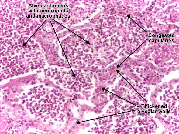
Leukocytic alveolitis is the 3rd phase of the lobar pneumonia. Alveolar lumens are filled with leukocytic (suppurative) exudate (neutrophils and macrophages, in order to remove the fibrin). Alveolar walls are thickened due to capillary congestion and edema. (H&E, ob. x20)
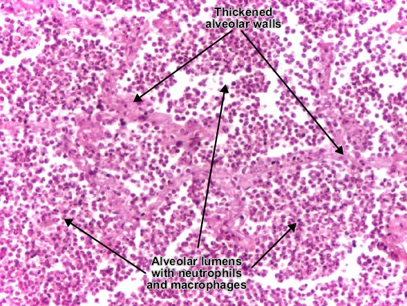
Lobar pneumonia, leukocytic alveolitis. (H&E, ob. x20)


Complication of pneumonia
1. delayed resolution
2. abscess formation
3. empyema
4. organization of exudates with fibrosis
5. Bactermia & Septicemia with infection to other organs eg. endocarditis ,pericarditis ,meningitis

I am very much happy to share to every viewers that is reading this,I want to inform the whole public of how I got help for my herpes, I wanted this since 6 months ago, I have also taken treatment from some doctor,few weeks back I came on the net to see if I will be able to get any information as to cure my herpes, on my search I saw various testimony of people who was helped by a great man called Dr Akhigbe and without any hesitation, I contacted him, I wrote to him and and he guided me, I asked him for solutions and he started the remedies for me and indeed 3 weeks after I started using the medicine, I was completely happy as it worked for me.I went to the hospital for check up and indeed I was declared negative from my disease, and I also waited again for two weeks and went back to another hospital for check up to be fully sure and to my great surprise I was still declared negative, and I decided to share this great opportunity to those people out there fighting this sickness, You can contact him now for your medicine to cure your diseases, contact his Email; drrealakhigbe@gmail.com or Whatsapp +2349010754824 website. hpps:drrealakhigbe.weebly.comDr Akhigbe also cure diseases like..
ReplyDeleteHIV, Herpes , Cancer, Chronic Disease, Asthma , Parkinson's disease, External infection, Als, progeria, common cold, multiple sclerosis disease, Nausea, Vomiting or Diarrhea, Heart Disease, meningitis, Esquizofrenia, Toxoplasmosis, Pulmonary Fibrosis,Diabetes, Kidney Disease, Lupus, Epilepsy, Stroke,Eczema, Erysipelas Eating Disorder, Back Pain. Osteoporosis etccontact him for your solution.